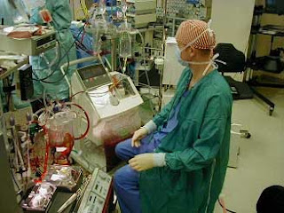
Weaning a patient off cardiopulmonary bypass (CPB) is an important step of cardiac surgical procedures. Emergence from CPB is a time for planned action and cooperation among the cardiac operating team. Returning heart and lungs to the circulation after CPB may represent a potential stress to the heart.
Most techniques, conduction and management of CPB are well standardized; however, separating patients from perfusion occasionally involves decisions based on empirical therapies. Weaning off CPB is a straightforward process that requires no particular measures other than reestablishing ventilation to the lungs and slowly turning the arterial pump off. In a number of cases however, weaning may be specially difficult, and in a few situations simply impossible.
Most techniques, conduction and management of CPB are well standardized; however, separating patients from perfusion occasionally involves decisions based on empirical therapies. Weaning off CPB is a straightforward process that requires no particular measures other than reestablishing ventilation to the lungs and slowly turning the arterial pump off. In a number of cases however, weaning may be specially difficult, and in a few situations simply impossible.
Separation of CPB may require special protocols, in accordance to the status of the patients, to their particular age group and the conduction of bypass. Weaning off small children after prolonged, difficult and complex operations may represent a challenge to the surgical team.
Weaning off CPB is always conducted in a coordinate fashion. The surgeon directs the weaning process with some input from anesthesia and perfusion. A skillful anesthesiologist usually shares the control of weaning with the surgeon. This allows the surgeon to direct full attention to the grafts position, valve function, security of suture lines and final gross hemostasis. When this arrangement fails or there is no free communication in the team, it is not unusual while separating a patient from bypass to observe a perfusionist retaining more volume into a hypotensive patient while the anesthesiologist administers a vasopressor, both ignoring the surgeon's observation that the heart is distending.
Inadequate weaning and separation from CPB can prolong recovery and increase morbidity and mortality.
PREPARING FOR WEANING
CPB is associated with various insults to normal physiology. These include anticoagulation, hemodilution, hypothermia, ischemic or chemical cardiac arrest, increased release of endogenous cathecolamine, vasopressin and other vasoactive substances, electrolyte disturbances, platelet activation, aggregation and destruction, and activation of complement and other plasma protein systems. These multiple interacting factors represent a number of potential routes for myocardial dysfunction or injury [1].
 Weaning process is initiated after adjustment of certain patient variables, such as temperature, tissue oxygenation, hematocrit level, acid-base and electrolytes status, and cardiac function. Requirements of analgesic, narcotic and paralyzing drugs usually increase during rewarming and the necessary adjustments are made with avoidance of myocardial and circulatory depressant agents [2].
 While most steps of weaning are standard and common to all cases, evaluation of cardiac function may disclose situations which will require specific measures such as the use of temporary pacemaker, inotropic or vasoactive drugs or mechanical support.
Temperature
Cardiopulmonary bypass is systematically accompanied by heat loss to the environment, even when hypothermia is not employed. Preparation for terminating CPB includes rewarming of patients to the normal or near normal temperature. Rewarming should be initiated as early as necessary so that its completion coincides with completion of the surgical procedure or soon afterwards. Patient temperature is monitored in at least two sites such as nasopharingeal, tympanic, esophageal, rectal or bladder. The most common combination is nasopharingeal and rectal temperatures.
As nasopharingeal temperature reaches 36.5 to 370C, rectal temperature is usually two to three degrees lower. A larger than four degrees gradient between the nasopharingeal and rectal temperatures is indicative of inadequate rewarming or increased vasoconstriction. In these situations there may occur a two to three degrees decrease in nasopharingeal temperature during sternal closure and transfer to intensive care unit, which may predispose the patients to unstable cardiac rhythm, shivering, and hypertension [3,4]. This afterdrop results from redistribution of heat within the body as the normal pulsatile blood flow opens up some relatively colder and constricted vascular beds [1,2].
A slow infusion of vasodilators (sodium nitroprusside) may provide a more homogenous rewarming and reduce the occurrence of significant temperature gradients. Pulsatile flow may promote the same results [5]. Warming blankets will not always be effective to correct the temperature drift, due to the increased peripheral constriction; small children may better benefit from warming blankets and ventilation with heated and humidified gases.
Hohn et cols [6] investigated 86 patients to draw the influence of warming the skin during weaning. Patients warmed through a water blanket and a blow of warm air to the head, in addition to the heat exchanger, had a better thermal balance and a lower blood loss, when compared to a control group.
Tissue oxygenation
Metabolic abnormalities are corrected before initiating the weaning process. Venous line carries true mixed venous blood, and venous oxygen saturation and PO2 are satisfactory indicators of tissue metabolism. Arterial PO2 and saturation best reflect the oxygenator performance.
Increased lactate production, decreased pH, and decreased mixed venous saturation and PO2 are indicators of inadequate tissue perfusion or oxygenation. A venous oxygen saturation of 75% and a minimum venous PO2 of 35mmHg are satisfactory to start weaning from CPB.
Hematocrit
Hemodilution is universally accepted as an important adjunct to cardiopulmonary bypass. Blood viscosity and oncotic pressure are reduced while tissue perfusion and oxygenation and cerebral flow are enhanced by the levels of hemodilution commonly used in clinical settings. Hematocrit values from 20 to 25% are usual with most perfusion protocols. By the end of rewarming, depending on renal function and the use of diuretics hematocrit may reach 24 to 30%.
Hearts with severe preoperative myocardial dysfunction will perform better immediately after termination of CPB with a hematocrit level above 34%. Red cells transfusion during rewarming may be necessary to adjust the hematocrit prior to discontinuing perfusion [7]. Under special circumstances or while perfusing small babies, a very low hematocrit can be corrected by transoperative ultrafiltration.
Acid base status
Regardless of the lowest temperature attained or the acid-base management protocol used (alpha or pH stat) during CPB, by the end of rewarming a pH of 7.4 and a PCO2 higher than 35 mmHg are mandatory to safely disconnect a patient from the pump.
Any degree of acidosis should promptly be corrected because it depresses myocardial contraction, diminishes the action of inotropes, and increases pulmonary vascular resistance.
Electrolytes
Potassium is the critical ion that may present acute changes during CPB, followed by calcium. Others ions rarely show significant changes and their correction is less demanding for the weaning to take place.
Hyperkalemia may result in atrioventricular block. Blood cardioplegia usually produces a higher potassium level at the end of perfusion. In the presence of a normal renal function, a mild hyperkalemia represented by a serum level of 6 mEq/L or less, will not require special treatment and resolve spontaneously. In the presence of a heart block or bradicardia, a more regular rhythm should be secured with temporary pacemaker wires. The hyperkalemia should be treated with insulin, glucose and furosemide [2,3].
Hypokalemia may predispose to atrial and ventricular arrhythmias, and shall be treated. During CPB it is preferable to administer potassium chloride in small frequent doses instead of continuous infusion. Doses of 5 mEq may be repeated after proper evaluation of serum levels.
Ionized calcium levels usually decrease during CPB and appears to recover fast thereafter. Administration of calcium chloride was widely employed in the past during separation from perfusion, because of its positive inotropic effect. Elevated serum calcium levels have been associated with increased vascular resistance on the peripheral, coronary, renal and cerebral microcirculation [1,8]. Calcium chloride administration has been associated with coronary arteries and mammary grafts spasms and is usually avoided in the revascularized patient. Some concern exists as to the potential role of an elevated serum calcium level in the aggravation of reperfusion injury [9].
Valvular and pediatric patients with a sluggish myocardial contraction frequently show some transient improvement after a small bolus (10 to 15mg/Kg) of calcium chloride immediately before discontinuing CPB.
Cardiac action
Several interacting factors of perfusion predispose the myocardium to injury and dysfunction. A certain amount of myocardial injury may be added after aortic unclamping, during the reperfusion phase. The period immediately before complete separation from bypass is critical; its duration is conditioned by myocardial recovery. Regardless of the myocardial protection strategy and method, even short periods of aortic cross-clamping can be followed by temporary functional depression.
Usually there is a mild and transient functional impairment shortly followed by resumption of effective cardiac action. Most hearts will benefit from a short period of supportive CPB, usually from 15 to 20 minutes for each hour of clamping. This is easily accomplished by properly timing surgery and rewarming [1,3,10].
When normal temperature is reached and preparations for weaning are completed, maximal myocardial recovery from the arrest period has usually been attained.
Cardiac function immediately prior to discontinuing perfusion is usually assessed by visual observation, electrocardiogram, ventricular filling pressures and afterload, and transesophageal echocardiography if available. Simple visual observation of heart action can provide valuable information on myocardial performance. Experienced teams can accurately predict the chances of immediate difficulties to terminate CPB by visually inspecting the heart action alone.
All clinical variables involved with cardiac performance (heart rate, preload and afterload, and contractility) are assessed in order to optimize cardiac output.
Cardiac rhythm and the adequacy of ventricular rate are evaluated by the electrocardiogram. Slow ventricular rates are adjusted by ventricular pacing; atrioventricular dissociation is corrected by atrioventricular sequential pacing.
Preload is evaluated by ventricular filling pressures. Left ventricular preload is inferred from left atrial mean pressure or pulmonary artery diastolic pressure; right atrial pressure reflects the preload conditions of right ventricle. During weaning preload is pump-dependent and can be adjusted by balancing blood volume between patient and oxygenator.
Ventricular afterload is evaluated by the status of peripheral vascular resistance. This is represented by the ratio between mean systemic arterial pressure and pump flow. Elevated peripheral resistance may require vasodilators. An occasional patient will present in a state of deep vasodilation and hypotension even when pump flow is adequate or elevated [11]. These will require a vasoconstrictor infusion to restore a normal peripheral resistance.
Transesophageal echocardiography (TEE) is useful to verify the adequacy of surgical repair in congenital and valvular cases; it can also offer valuable information on ventricular volumes and the quality of myocardial contractility [12].
Ninomiya et cols [13] assessed continuous transesophageal echocardiography monitoring during weaning from CPB in 41 children. They measured left ventricular ejection fraction, wall motion and end-diastolic volume. In the presence of severe heart failure, the authors could adjust drugs and mechanical support oriented by the TEE information. Shankar et cols [14] found epicardial ultrasound examination of paramount importance to detect less than perfect coronary translocations after Jatene's operation.
TERMINATION OF CPB
After adjusting cardiac rhythm and rate, preload and systemic arterial resistance, the assessment of cardiac function immediately before terminating CPB allows the patients to be classified into 3 groups. The proportion of patients on each group will depend on case distribution. According to our retrospective experience [15], a general surgical service dealing with the broadest spectrum of patients, which includes elderly, reoperations, emergencies and neonates will result in an approximate 70% of patients in group A, 25% in group B and 5% in group C.
Group A: Patients that will obviously offer no difficulty to disconnect from perfusion. For these patients, after reestablishing ventilation to the lungs, pump flow can be gradually reduced while venous return to the oxygenator is decreased until bypass is minimal. Arterial pump is stopped and venous line is clamped. Final adjustment of cardiac performance is made off pump, by slowly administering residual volume from the oxygenator until ideal preload is attained. These patients maintain an adequate cardiac output, as can be confirmed by normal atrial and arterial pressures, arterial and venous blood gases and pH and adequate spontaneous diuresis.
Most teams will administer a slow infusion of an inotrope (dopamine or dobutamine) or, less frequently a vasodilator, based only on "routine" protocol or "past" experience. This infusion is usually discontinued as the patient arrives to the intensive care area or is maintained for a few hours thereafter.
Group B: Patients with a mild to moderate degree of cardiac dysfunction that will require some support to disconnect from the pump. This support may be physiological (Starling law) or pharmacological (inotropes, vasodilators or both). Some patients in this group can benefit from intra-aortic balloon pumping [16,17].
Group B patients require a more elaborated protocol for CPB termination. Final preparations are made on partial bypass.
Before turning pump off all clinical determinants of cardiac performance are evaluated and adjusted, in order to optimize cardiac output. Blood volume is adjusted according to left atrial or pulmonary artery pressures and inotropes are commenced. Peripheral resistance is estimated and vasodilators or constrictors are instituted as required. After the drugs effectiveness is assessed, pump flow is decreased in small increments while venous return in proportionately adjusted to maintain a constant filling pressure. Arterial pump is stopped and venous line is clamped.
Most patients in group B will perform as well as group A patients. Some patients may have to return to pump for better adjustment of drugs, or to have an intra-aortic balloon inserted if a marginal cardiac output is present, as demonstrated by atrial and arterial pressures, arterial and venous blood gases and pH, and spontaneous diuresis.
Children with preoperative high pulmonary blood flow, children after a heart transplant, and some adults with long standing congestive heart failure may present with pulmonary hypertension that precludes successful weaning. Inhalation of nitric oxide (NO) has been demonstrated as dramatically improving cardiac output and allowing a smooth discontinuance of CPB [18, 19].
Bauer et cols [20] evaluated the efficacy of prostaglandin E1 as a poweful adjunt to wean difficult transplanted children with right ventricular failure.
The association of epinephrin in a slow infusion and nitroprusside or another vasodilator drug, possibly represents the strongest available stimulus to improve myocardial contractility.
A recently introduced inotrope (enoximone) is under evaluation to provide phrmacological support during weaning of patients with severe ventricular dysfunction [21,22].
An occasional patient in group B will not tolerate CPB termination even after a few trials. These few exceptions turn into group C patients.
Group C: Patients with severe cardiac dysfunction that will prove difficult to be removed from CPB, despite physiologic and pharmacological support. For these patients CPB will have to be prolonged. A few hours of circulatory assistance and intensive inotropic and vasodilator drugs therapy may turn some of these patients into group B. The remaining patients are candidates to a form of total circulatory mechanical support (if available) or they will not likely survive disconnection from pump [23,24,25].
Group C patients are by definition the hardest cases to manage. A few of these patients by the end of rewarming will have minimal or no cardiac activity which precludes any trial of disconnection from pump. The remaining patients may be given a short trial off pump after optimization of preload, afterload and contractility by a criterious combination of inotropes and vasoactive agents. Some of these patients will tolerate CPB removal, under maximal physiological and pharmacological support, and a few in the group may be further improved by an intra-aortic balloon pump. The patients with minimal cardiac activity and those in whom the trial off pump was unsuccessful are temporarily maintained on cardiac support with the heart-lung machine. A few hours on pump support may be a sufficient rest period to allow recovery of cardiac function and removal of CPB support in a small number of cases. For the others, a decision has to be made as to either advance to a mechanical device for prolonged support or terminate the efforts to recover cardiac action.
Children supported by full veno-arterial extracorporeal membrane oxygenation (ECMO) post cardiotomy, have a poor long term survival rate [26] when compared with children managed with centrifugal ventricular assist devices [27].
CONCLUSION
Weaning and disconnecting CPB is a team effort and requires clear planning and integrated performance. Despite being a simple procedure, interrupting perfusion can be elaborated, extremely difficult or virtually impossible. Resources for prolonged supportive bypass and mechanical devices shall be made available and their application to the difficult situations should be a part of CPB protocols.
SUMMARY
Weaning off cardiopulmonary bypass is a simple process; however, it may occasionally prove very difficult and sometimes virtually impossible. Preparation for weaning includes the adjustment of several patient related variables such as temperatures, tissue oxygenation, hematocrit, pH, electrolytes, and cardiac rhythm and rate. Cardiac action as routinely assessed will allow patients to be classified into 3 groups. Group A includes patients which will obviously offer no difficulties to remove from CPB. Group B comprises patients with mild to moderate dysfunction; they will require some physiological (Starling law) or pharmacological (inotropes, vasoactive drugs) support to be disconnected from pump. Group C are the patients with poor or no cardiac action, which will require either an aggressive pharmacological support or prolonged mechanical support as alternatives to sustain life.< p align=justify> Resources for prolonged supportive bypass and mechanical devices shall be available and their application to difficult situations should be part of CPB protocols.

